Diagnostic imaging: What’s the difference between X-ray, ultrasound and MRI?
X-rays, ultrasound and MRIs are all types of diagnostic imaging tools that help members of the medical profession see what's going on in the inside, but how they work and what each one tells us is very different. Here we explain more...
What’s an x-ray?
X-raying uses a small amount of radiation to quickly capture a single image of your anatomy to assess fractures or dislocations or disease (bone degeneration, infections or tumours). When the rays pass through the body they are absorbed by the different tissues. The denser the tissue, the whiter the image, as more x-rays are absorbed and so don’t pass through, which is why lungs are black and bones are white. This is why it is difficult to image tissue behind bones as that’s already absorbed the majority of the rays.
What’s ultrasound?
Ultrasound uses sound waves instead of radio waves to produce a picture within your body in real-time, and can assess the condition of organs and soft issues that Xrays can’t. Ultrasound therapy is also used by physios or occupational therapists to treat pain, reduce swelling and inflammation and to promote tissue healing.
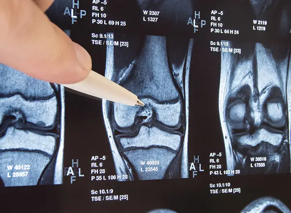
What’s an MRI scan?
An MRI (magnetic resonance imaging) combines a powerful magnetic field with an advanced computer system and radio waves, and is used for imaging organs, soft tissue and internal structures; showing tissue difference between normal and abnormal; and imaging without radiation. MRI is generally recommended when doctors need to see a more detailed view of what’s happening inside or behind bony structures or air-filled organs, such as the lungs that the other diagnostic tools can’t get to.
- How do you tell the difference between a soft tissue injury and a stress fracture?
- What’s the difference between shin splints and stress fractures?
 220 Triathlon Team
Journalists, reviewers, coaches and athletes
220 Triathlon Team
Journalists, reviewers, coaches and athletes